Lung cancer is one of the most important deadly diseases in the world. The recent estimates provided by the World Health Organization (WHO) says that around 7.6 million deaths worldwide per year due to lung cancer. Moreover, humanity due to cancer is supposed to continue rising, to become around 17 million worldwide in 2030.
Discovering lung cancer in the early stage is the only method for its cure. Different methods are available for diagnosis lung cancer, namely, MRI, isotope, X-ray and CT. X-ray chest radiography and Computer Tomography (CT) are the two familiar anatomic imaging modalities that are regularly used in the recognition of different lung diseases. CT images are used by physicians and radiologists to identify and recognize the presence of diseases, directly visualize the morphologic extents of diseases, describe the patterns and severity of diseases, and measure the clinical course of diseases and response to therapy.
The volumetric CT technique has introduced spiral scans that shorten the scan time and, when used in thoracic imaging, reduce the artefacts caused by partial volume effects, cardiac motion, and unequal respiratory cycles. As the progress of CT technology, the high-resolution CT test has happen to the imaging modality of choice for the recognition and identification of lung diseases. Even though High-Resolution Computed Tomography (HRCT) recommends images of the lung with progressively more improved anatomic resolution, visual interpretation or evaluation of a large number CT image slices remains as a difficult task.
Abstract
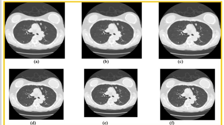
Lung cancer is one of the main reasons for death in the world among both men and women, with an impressive rate of about five million deadly cases per year. Computed Tomography (CT) scan can provide valuable information in the diagnosis of lung diseases. The main objective of this work is to detect the cancerous lung nodules from the given input lung image and to classify lung cancer and its severity. To detect the location of the cancerous lung nodules, this work uses novel Deep learning methods.
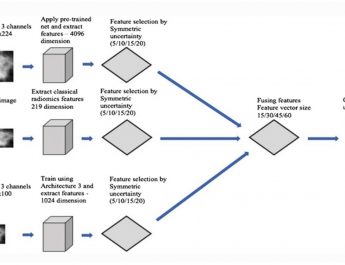
This work uses best feature extraction techniques such as Histogram of oriented Gradients (HoG), wavelet transform-based features, Local Binary Pattern (LBP), Scale Invariant Feature Transform (SIFT) and Zernike Moment. After extracting texture, geometric, volumetric and intensity features, Fuzzy Particle Swarm Optimization (FPSO) algorithm is applied for selecting the best feature. Finally, these features are classified using Deep learning. A novel FPSOCNN reduces computational complexity of CNN. An additional valuation is performed on another dataset coming from Arthi Scan Hospital which is a real-time data set. From the experimental results, it is shown that novel FPSOCNN performs better than other techniques.
A. Asuntha & Andy Srinivasan