Paul Andersen explains the structures and functions of seventeen major parts of the brain in this video. He begins with a quick discussion of brain evolution and ends with a review of the major parts presented inside the brainstem, cerebellum, thalamus, and cerebrum.
“It’s Mr. Andersen and in this video, I’m going to talk about the brain, structure, and function. Remember! The structure is what it’s made up of. And Function is what does it do. We sometimes refer to this as the anatomy or structure and physiology or the function. And so the cool thing is that we’re going to go through seventeen different structures in the brain, kind of layout the basic plan of the brain. But you are using your brain to process it. “

And so what type of organisms have brains?
It’s animals. Animals use nerves. They have muscles to move around. And so they have to organize that movement. And so they use a brain. And so if we look at the two basic body plans of animals, some are radially symmetrical.

In other words, they’re built around almost a tire. And then some are bilaterally symmetrical. In other words a tiger you could draw a line right down the middle. There’s going to be a clear right side and a left side. There’s going to be a clear front and end. And as we became bilaterally symmetrical we had to organize that movement.

And so this is a simple animal body plan. And so this animal is going to move towards the right. And as it does so it has to take in information. We call that sensory information using neurons. And so right now you’re taking in sensory information from your eyes, from your ears. And then inside your brain, you’re going to integrate that information. You’re going to make sense of it. And then you’re going to figure out what you want to do. How you’re going to act dependent upon that. And so then we have this loop of motor neurons out. Or motor nerves. And so this loop in simple animals is also important in understanding how our brain works.

But if we look at these real primitive brains we find that they have a real common structure. They have these four humps. And we call those, well the first one is not a hump, but the spinal cord. We then have the hindbrain, the midbrain and then we finally have the forebrain. And we find this consistent throughout all animals.
And if we look at something like a shark, it pretty much looks just like that primitive brain. You can see down here we’ve got the spinal cord that’s bringing information in. We then have the hindbrain, the midbrain, and the forebrain.

And so one thing you should remember is that the closer we are to that spinal cord, the more basic the functions are. And so we’re right down in this hindbrain. It’s going to be basically keeping the heart beating. Keep the circulation going. Digestion in the shark. But when a shark decides to attack you or it has some kind of an emotional response, that’s going to be way up here in the forebrain.
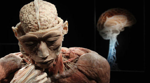
Now if we look at you when you were really little, when you were an embryo, you had a brain that looked very similar. You had a spinal cord. You then had a hindbrain. You had midbrain. And then you had a forebrain. But during development that brain changes radically. And so this is what an adult brain looks like. So we still see that spinal cord. We then have the hindbrain. We have the midbrain. But look how large that forebrain is going to be. So that’s where all of those emotions and memories and all of that thinking, we generally attribute to the brain is going to be in the forebrain.

And so let’s get to the actual anatomy. And so there are going to be 17 parts that we’re going to go through. So you should always be thinking what’s the name of the structure? Where is it? And then what’s the function, what does it do?
So if we look at a basic brain plan we find these four things jump out right away. We’re going to see the brainstem. We then see a cerebellum on the back of the brain So again to get yourself oriented right the eyes are going to be right up here. So this would be towards the back of the head. So that’s going to be the cerebellum. We then have the area of the thalamus hypothalamus. And then finally we have the cerebrum which is going to be that dominant upper portion of the brain.

And so let’s begin with the brainstem. The brainstem is broken down into three individual structures. So if we start at the bottom we’ve got the medulla oblongata, the pons and then we finally have the midbrain. And so those three things, medulla oblongata, pons, and midbrain make up what we call the brainstem. So that the structure. What’s the function? Well, it really does two things. The first thing it’s going to do is these more basic needs. It’s going to keep yourself breathing, keep circulation going, digestion, swallowing. All of that is going to be controlled by the brainstem. If there’s any damage to the brainstem it’s going to be catastrophic.

What else does it do? Then we have information coming in. So we have sensory information, just like that worm did, coming up to the brain. And then we have motor nerves going out. And so the brainstem is important in routing that information and filtering that information, sending it where it needs to go.
What’s behind that? We have the cerebellum. The cerebellum and the function of that is motor control. So as you do sports, for example, it’s the cerebellum that’s giving you that coordination. And it also gives you motor memory. So as you learn to ride a bicycle and you remember how to ride a bicycle that’s going to be thanks to your cerebellum.

If we keep moving up we now have the thalamus. The thalamus again sits right on top of the brainstem. And so the best analogy I could come up with is a router. It’s basically sorting data and sending it where it needs to go.
If we were to look below that there’s a little structure here that’s incredibly important. It’s called the hypothalamus. That’s going to be really right above the roof of your mouth. What is that accountable for? It’s homeostasis. So it’s maintaining body temperature. It’s maintaining osmolarity. All of that stuff is contained right up in the hypothalamus. Also important in circadian rhythms.
And then if we look right below that you can see a little gland hanging out. And one-half of that pituitary gland, the posterior pituitary, is technically part of the brain. And it’s important in basically sending off hormones. And so there are nerves that flow into that pituitary and it’s sending out things like an antidiuretic hormone. That keeps your water balance the same. Oxytocin would be another important hormone that comes out of there.

If we keep moving up then we get to the level of the cerebrum. What’s the function of the cerebrum? That is integration. So what we’re doing is making sense of all of that data that comes in. Now, what makes up that cerebrum is going to be all these neurons. There are tons of neurons that are connected together. Billions of neurons. And billions and billions of synapses or connections between these neurons. And that’s where we’re making sense of information as it comes in.
What we find is as you look at those images your brain is integrating. It’s making sense of all that information. And it used to be a black box. We didn’t know really what was going on. But now we can use technology as a functional MRI. A functional magnetic resonance imaging. And what we’re looking at here is a brain in action. So this same study was done on females. And what they would show them is something neutral, like a brick wall. And then a kitten. And then something like dirt. And then something like puppies. And so what we’re seeing is as those images are switching back and forth we can start to see where blood is flowing around in the brain and we can start to figure out what the different parts of the brain actually do. We’re able to figure out their function.
So when we’re looking at the cerebrum every picture that I’ve shown you is from the side. So the eye is up here. But if we were to rotate that 90 degrees now were looking at it head-on, we’ll find that there are two hemispheres. There’s going to be a right and a left hemisphere. Now they are connected in the middle using something called the corpus callosum. So that’s a connection of nerves in between the two hemispheres. And we do tend to show lateralization. There are going to be certain things that we put kind of on the left side of our brain, like mathematical reasoning and logic. And things that we put on the right side like facial recognition. Now, this is plastic. In other words, we can move these functions back and forth. And you can even have a radical hemispherectomy, where you’re cutting one of these out and you still have a functioning brain.

Now if we were to go right below the corpus callosum we get into this area called the basal ganglia. And it’s made up of a bunch of nuclei. What are nuclei? Or what is a nucleus in a brain? It’s basically a bunch of neurons that are right next to each other that have the same function. And so all of these nuclei together make up what’s called the basal ganglia. And you can see this would be the corpus callosum, connecting it together as well. So this is below the cerebral cortex.
What’s the function of that? Well, scientists have been able to figure out there is this complex interaction of inhibition and excitatory response between these neurons. And basically it controls a lot of our motor control. And if you have somebody who has Parkinson’s disease then we’re having problems in this basal ganglia area.
As we move farther up the brain we eventually get to the cerebral cortex. And that’s going to make up about 80 percent of the brain. So it’s most of the brain itself. And it’s broken apart into these four lobes. And so if we start in the front of the brain we have what’s called the frontal lobe. What’s the function of that? It’s mostly executive function. So it’s kind of like the boss of your brain. It’s emotional control up there. And if we have people who have damage to that frontal lobe they have really huge emotional swings.

As we move back towards the back of the brain we get to the parietal lobe. What’s the function of that? It basically is a sensation. It’s you dealing with and reacting to your environment. So we have a lot of neurons coming in here from sensory input. As we move to the back we have the occipital lobe. The function of that is the vision, primarily vision. And then we move on to the side. We have what is called the temporal lobe. The temporal lobe is going to be important in language. It’s important in hearing. It’s also important in memory. We have a lot of memories in there. And so each of these lobes has different functions that are associated with it. And hopefully, those little icons help you remember those functions.
Now if we were to go inside the parietal zone we’d find a really important part here. It’s called the somatosensory cortex. And that’s where sensory information is coming into the brain. And then on the other side of the lobe, we have what’s called the motor cortex. And so going way back to that worm, we have information coming in, sensory information. And then we have motor output coming out. And so that’s going to be a point of integration where we get information in. Decide what we want to do with it. And then send that message back out.

Now if we were to look at that somatosensory cortex and map it along the cerebral cortex, we would find that we dedicate huge amounts of that brain surface area to things like your fingers, your tongue, your lips. In other words, we have way more neurons and way more sensory information coming in from your fingers as opposed to, for example, your back. We don’t have as much of it dedicated to that on the backside.
We could also functional MRIs and then even an operation to figure out where a lot of these things are located, like speech and smell and hearing. But over the future, we’re going to get really really good at figuring out specifically what are all of the different parts of the brain. What are the nuclei? What do they do? And even mapping it down to the level of the neuron.